

TEER Measurement
EVOM is the leading range of TEER meters. They provide a reliable method to confirm the integrity and permeability of the monolayer.

WPI pioneered the production of TEER (Trans Epithelial Electrical Resistance) measurement equipment in the 1980s and is still innovating new equipment for cell researchers.
You've placed your trust in our TEER measurement line for nearly 40 years as the premiere innovator and manufacturer of EVOM™ meters, REMS, and Millicell ERS 2. Learn more about the gold standard in TEER technology that has been cited in over 16,000 peer-reviewed research articles.
Transepithelial Electrical Resistance (TEER) qualitatively measures cellular monolayer health and quantitatively evaluates cellular confluence by measuring the electrical resistance across a cell monolayer. It is commonly used to assess the integrity and permeability of cellular barriers, such as epithelial and endothelial cell layers in cell culture plates. TEER has proven to be a valuable tool in various fields, including drug absorption studies, tissue engineering, and disease modeling. TEER measurement is non-invasive so can be used to monitor live cells during their various stages of growth and differentiation.
TEER Measurement Equipment
EVOM TEER Measurement Basic Working Principle
What is TEER?
TEER is a measure of the resistance to the flow of ions across a cell layer grown on a porous membrane. It reflects the tightness of the cell junctions and the barrier function of the cell layer. Higher TEER values indicate tighter junctions and a less permeable barrier, while lower TEER values suggest looser junctions and a more permeable barrier. When culturing cells in a well plates, TEER can be used to determine cellular confluence. When the TEER value rises steeply, confluence has been achieved and then barrier studies may begin.
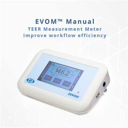
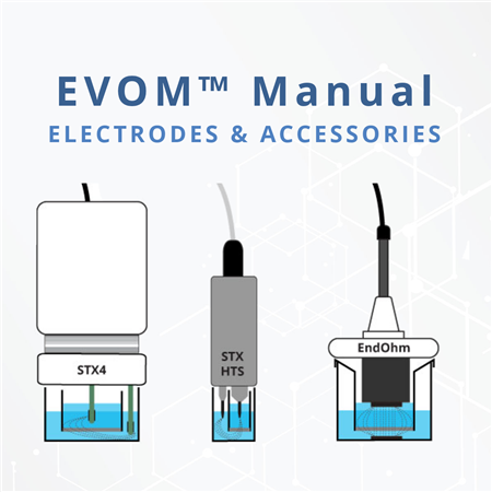
TEER is typically measured using a probe with a pair of electrodes placed on either side of the cell monolayer, one in the apical compartment (above the cells) and one in the basolateral compartment (below the cells). An electronic instrument like the EVOM™ Auto or EVOM™ Manual applies a small, defined voltage across the electrodes and measures the resulting current, from which the resistance can be calculated.
The TEER value is calculated using Ohm's Law (R = V/I), where R is resistance, V is voltage, and I is current. It is usually reported in units of ohms times square centimeters (Ω·cm²) to account for the area of the cell monolayer.
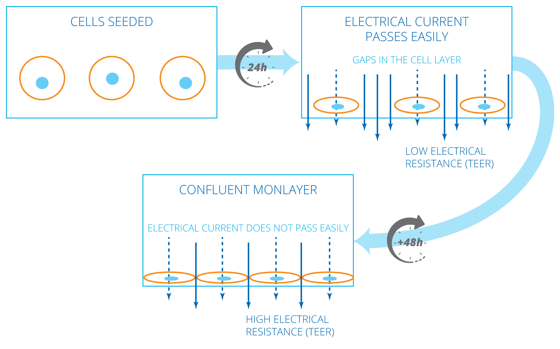
Initially, 24 hours after cell seeding transwell, TEER values are generally low, because the current passes can pass easily between the cells. Over time, the cells multiply and start covering the gaps. Finally, a confluent cellular monolayer is formed. At that point, the permeable membrane is fully covered with cells and does not allow easy passage of electrical current. This results in a high TEER value.
TEER Measurement of Leaky & Tight Cell Types
TEER values of confluent cellular monolayers can vary depending on the cell type. Monolayers of certain cell types (e.g., cell type A), which normally show low TEER values, generally have relatively leaky tight junctions. Monolayers of other cell types (e.g., cell type B) show high TEER values, and these cell types are known to have tight junctions. Ions and molecules are known to pass rather easily across leaky cellular layers as compared to tighter cellular layers. The presence of more transcellular ion channels on cells can further allow easier flow of ions or electrical current through the transcellular pathway, which can additionally lower TEER values.

Cell type A allows greater amounts of current and ions to pass between the cells and yields a low TEER value. With its tighter junctions, cell monolayers of type B cells will show a higher TEER value. While both monolayers are confluent, the TEER resistance values can be markedly different based on the nature of the cells themselves.
TEER Value Calculation
TEER is a normalized value of resistance per 1 square centimeter of unit area. To compute TEER, multiply the measured resistance by the surface area listed below. For example, a 6.6 mm insert measures 1707 Ω, the TEER is 1707 Ω * 0.331 or 505 Ω.
- 6 well plate (24 mm inserts) 4.53 cm2
- 12 well plate (12 mm inserts) 1.13 cm2
- 24 well plate (6.5 mm inserts) 0.3316 cm2
- 96 well plate (4.3 mm inserts) 0.145 cm2
TEER Measurement In Epithelial Cell Research
In epithelial cell research, there are different types of cells which are typically cultured. Epithelial tissue lines the surface and cavities of an organ. Epithelium includes your skin tissue, but it also lines the alimentary canal, organs and blood vessels. Epithelial cell cultures may be used for drug discovery or other applications that explore the transport or diffusion of a substance through an epithelial monolayer. Such a tissue research study requires a confluent monolayer that completely covers the culture substrate it is grown on.
The in vitro cell culture models of human endothelial and epithelial monolayers are considered as reliable models of the in vivo environments and as such are used for drug toxicity and transport studies. Findings of the in vitro models are also translated to ascertain the metabolic and physiological functions of a particular pharmacological entity. The most commonly used endothelial/ epithelilal in vitro models are:
- Blood-brain barrier (BBB) model
- Gastrointestinal tract (GIT) model
- Pulmonary models (including viral infection model, such as COVID-19)
These are used for understanding the absorption and transport of drugs, as well as associated cytotoxicities in the organs like the central nervous system, intestine, and lungs. These models are either comprised of primary cells or established cell lines. For utilization of these models, the most important feature is to ascertain the capability of cells to form necessary intercellular junctions. Before proceeding with particular drug cytotoxicity or transport, the formation of cellular junctions in these in vitro models is confirmed through a variety of methodologies. This include the permeability of the established barrier to compounds like sucrose having radiolabeled carbon, and lower molecular weight paracellular tracers including inulin, mannitol and albumin.
Besides all the above described methodologies for determining the integrity of an in vitro endothelial/epithelial barriers, transepithelial/transendothelial electrical resistance (TEER) measurements are considered more precise and highly quantitative in nature. More importantly, the TEER measurements are faster, inflicting minimal to no damage to the cells and with nothing being added externally, as the addition of any tracer or radiolabeled compound can potentially an potentially affect cellular physiology of monolayers.
The EVOM™ meters by WPI are a unique TEER measurement systems that are considered highly reliable for evaluating the integrity of in vitro epithelial barrier models including blood-brain barrier, gastrointestinal tract, and pulmonary model.
What are the Benefits of TEER Measurement?
Assessing Epithelial Barrier FunctionTEER allows researchers to evaluate the integrity and functionality of endothelial and epithelial barriers, such as the gastrointestinal, respiratory, and blood-brain barriers. By measuring the resistance across these barriers electrically, TEER provides quantitative data on the tightness and permeability of these barriers. This information is crucial for understanding drug absorption, transport mechanisms, and the effects of various compounds on barrier function. |
|
Drug Absorption StudiesTEER is extensively used in drug absorption studies to assess the permeability of drugs across tissue barriers. By measuring TEER before and after drug exposure, researchers can determine the impact of drugs on barrier integrity and evaluate their potential for absorption into a given tissue. This information aids in drug formulation, optimization, and predicting drug bioavailability. |
|
Tissue Engineering and Cell CultureTEER is a valuable tool in tissue engineering and cell culture studies. It helps researchers assess the formation and functionality of epithelial cell layers, mimicking in vivo conditions. TEER measurements can guide the optimization of culture conditions, scaffold design, and cell differentiation protocols. Additionally, TEER can be used to monitor the barrier function of tissue-engineered constructs over time, providing insights into their long-term viability and functionality. |
|
Disease ModelingTEER is an invaluable tool for disease modeling, particularly for studying disorders affecting endothelial and epithelial barriers tissues. Researchers can use TEER to investigate the impact of diseases, pathogens, or toxins on barrier integrity and function. This enables a better understanding of disease mechanisms, identification of potential therapeutic targets, and evaluation of drug efficacy in disease models.
|
|
Quality Control in Cell-Based Assays and Cell TherapiesTEER serves as a quality control measure in cell-based assays involving endothelial and epithelial cells. It ensures the consistency and reliability of experimental results by confirming the formation of tight junctions and functional epithelial barriers. TEER measurements can help identify potential issues with cell culture conditions, cell quality, or experimental protocols, ensuring the validity of research findings. Furthermore, TEER is now being implemented as a quantitative quality control measure for certain cell therapies. |
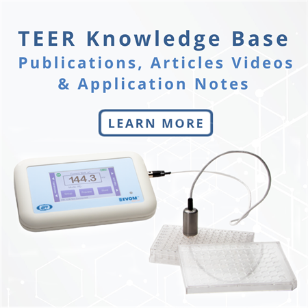
TEER Measurement Knowledgebase
Visit our knowledgebase for more technical information
A good overview of TEER measurement techniques is given in the paper below.
Srinivasan B, Kolli AR, Esch MB, Abaci HE, Shuler ML, Hickman JJ. TEER Measurement Techniques for In Vitro Barrier Model Systems. Journal of Laboratory Automation. 2015;20(2):107-126. doi:10.1177/2211068214561025

EVOM™ Auto
EVOM™ Auto automates measurements of TEER epithelial or endothelial monolayers...

Multi Well Plates And Cell Culture Inserts
WPI offer a range of multi-well plates, HTS plates and cell culture inserts availabl...

REMS
REMS AutoSampler automates measurements of TEER epithelial or endothelial monolayers...
EVA-MT-03-01
EVOM Auto For TEER Measurement in 96 HTS Plate
System includes:
- EVOM™ Auto Autosampler
- HTS Electrode Array for 96 HTS Plates
- Interface Unit and Cable
- Control iPad with software
EVA-MT-03-02
EVOM Auto For TEER Measurement in Corning 24 HTS Plates
System includes:
- EVOM™ Auto Autosampler
- HTS Electrode Array for Corning 24 HTS Plates
- Interface Unit and Cable
- Control iPad with software
EVA-MT-03-03
EVOM Auto For TEER Measurement in Millipore 24 HTS Plates
System includes:
- EVOM™ Auto Autosampler
- HTS Electrode Array for Millipore 24 HTS Plates
- Interface Unit and Cable
- Control iPad with software
EVM-AC-03-03
EVOM™ Warming Plate - Minimize the effect of temperature fluctuation by taking...





Request
Catalogue
Chat
Print