

MCS
MOM Computer System and Software- Overview
- Specifications
- Accessories
- Citations
- Related Products
Overview
There are 1 images available to view - click to enlarge and scroll through the product gallery.
MCS DataSheet
/ Download as PDF
Features
- Large-scale, high-resolution, deep-tissue mapping
- Multispectral, high-speed, functional optical imaging
- Photostimulation while imaging through the same optical pathway ("photostimaging")
- Two-photon microscopy and concurrent electrophysiology with computer-controlled placement of electrodes by Sutter micromanipulators
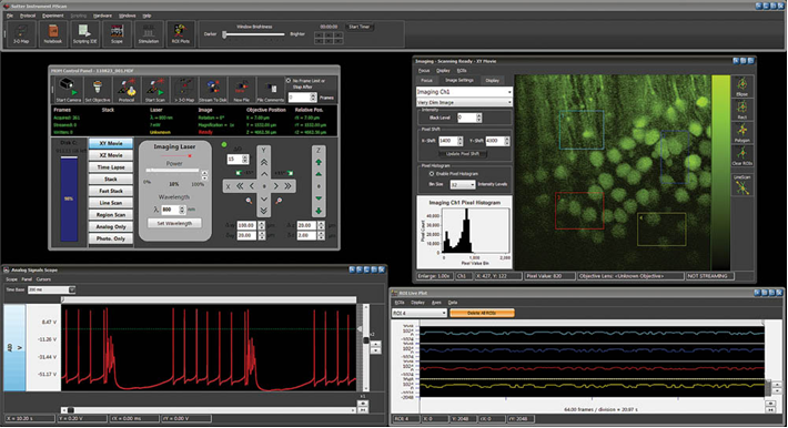
The MOM Computer System (MCS) includes the software package MScan. This program has been designed to seamlessly control two-photon imaging using conventional or resonant scanners while incorporating photostimulation and electrophysiology. While designed exclusively for use with the MOM microscope, it is also compatible with other two-photon platforms. MCS2.0 is designed to take on complex experiments in deep-tissue intravital imaging. Its intuitive user interface is easy to use. The MCS package and the MOM together form a formidable tool to understand the most complex issues in neuroscience, immunology or oncology. Importantly, you will find in MCS the same standard of technical excellence that is the hallmark of all Sutter Instruments products.
The MScan software has been developed to simplify the many tasks inherent in complicated imaging experiments. MScan is extensively multithreaded to take advantage of multicore processors. The newest release, MScan2.0 supports resonant or conventional scanning. MScan is multiuser based to facilitate sharing of a MOM microscope with MCS among experimenters. Experimenters can then send their data to other workstations for analysis. The MCS analysis program MView is available for free download on the Sutter Instrument website.
MCS includes a windows 7 workstation, National Instruments data acquisition boards, a firewire CCD camera and a USB controlled MPC-200. The National Instruments boards included are a PCI-6110 board for imaging, a PCIe-6353 board for control of imaging and photostimulation laser power and a PCIe-6321 board for electrophysiology. The package is a turnkey system as all data acquisition boards and software come installed within the workstation.
An important feature in MScan is the ability to do bidirectional frame scanning with sub-pixel line offset adjustment. Conventional two-photon frame scanning has involved unidirectional scanning. In these scenarios, data is only recorded when sweeping in one direction across the sample. To increase the rate of data acquisition, it is then necessary to steer the laser beam back to the origin of the scan as quickly as possible to begin the subsequent line. As galvanometric scanners are most taxed and most likely to be damaged during these high-frequency movements, bidirectional scanning both increases the speed at which frames can be recorded and decreases the likelihood of damage to expensive galvanometers.
Technical Information
IMAGING MODES
|
IMAGING FEATURES
|
Specifications
TECHNICAL SPECIFICATIONS
Dimensions (subject to change)
CPU
20.5in x 8.25in x 20in | 52cm x 21cm x 51cm
Monitor
26in x 10in x 19in | 67cm x 24cm x 48cm
Weight (subject to change)
CPU
31 lbs | 14 kg
Monitor
15 lbs | 7 kg
Electrical
115/ 230 Volts
50/ 60 Hertz power line
RoHS compliant
MCS SYSTEM
Catalog Number and Description
MOM-MCS*
System includes preinstalled MCS2.0 software, Windows 7 workstation, data acquisition boards, Firewire CCD camera
* Must be used with MOM systems equipped with MOM-DAQ, MPC-200 and ROE-200
MCS COMPONENTS
Catalog Number and Description
MOM-DAQ
MOM data acquisition system (includes NI 6110E PCI board)
MPC-200-ROE
Includes MPC-200 controller and ROE-200
NOTE: The MOM-MCS communicates with the MPC-200 controller via the USB port for control of X, Y and Z movement. It is not compatible with the MP-285 controller. Please contact Sutter for more information.
Accessories
Citations
Anwar, H., & Roome, C. (2014). Dendritic diameters affect the spatial variability of intracellular calcium dynamics in computer models. Name: Frontiers in …. Retrieved from https://journal.frontiersin.org/Journal/10.3389/fncel.2014.00168/pdf
Eggermann, E., Kremer, Y., Crochet, S., & Petersen, C. (2014). Cholinergic Signals in Mouse Barrel Cortex during Active Whisker Sensing. Cell Reports. Retrieved from https://www.sciencedirect.com/science/article/pii/S2211124714009590
Letourneur, A., & Chen, V. (2014). A method for longitudinal, transcranial imaging of blood flow and remodeling of the cerebral vasculature in postnatal mice. Physiological …. Retrieved from https://physreports.physiology.org/content/2/12/e12238.abstract
Park, J., Frantz, M., Kast, R., & Chapman, K. (2014). Nogo Receptor 1 Limits Tactile Task Performance Independent of Basal Anatomical Plasticity. PloS One. Retrieved from https://dx.plos.org/10.1371/journal.pone.0112678
Paukert, M., Agarwal, A., Cha, J., & Doze, V. (2014). Norepinephrine controls astroglial responsiveness to local circuit activity. Neuron. Retrieved from https://www.sciencedirect.com/science/article/pii/S0896627314003535
Roome, C., & Kuhn, B. (2014). Chronic cranial window with access port for repeated cellular manipulations, drug application, and electrophysiology. Frontiers in Cellular Neuroscience. Retrieved from https://www.ncbi.nlm.nih.gov/pmc/articles/PMC4227473/
Seemann, K., & Kuhn, B. (2014). Multi-photon excited luminescence of magnetic FePt core-shell nanoparticles. Biomedical Optics Express. Retrieved from https://www.opticsinfobase.org/boe/fulltext.cfm?uri=boe-5-7-2446&id=294401
Suzuki, N., Tang, C., & Bekkers, J. (2014). Persistent barrage firing in cortical interneurons can be induced in vivo and may be important for the suppression of epileptiform activity. Frontiers in Cellular …. Retrieved from https://www.ncbi.nlm.nih.gov/pmc/articles/PMC3952511/
Tada, M., & Takeuchi, A. (2014). A highly sensitive fluorescent indicator dye for calcium imaging of neural activity in vitro and in vivo. European Journal of …. Retrieved from https://onlinelibrary.wiley.com/doi/10.1111/ejn.12476/full


Request
Catalogue
Chat
Print