

TBI-0310 Head Impactor
TBI-0310 Head Impactor
A head trauma contusion device designed for medical research using rats and mice
- Overview
- Specifications
- Accessories
- Citations
- Related Products
Overview
There are 1 images available to view - click to enlarge and scroll through the product gallery.
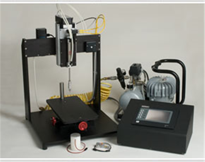
This system provides researchers specializing in traumatic brain injury a unique instrument to evaluate the mechanisms underlying brain trauma.
The instrument enables the application of standard contusion injuries to the brains of small rodents.
The amount of cortical depression is user-selectable between 0 and 3.5 mm (0.01 mm increments).
The target velocity of the impacting tip is electronically monitored and is user-selectable between 1.5 and 6.0 m/sec (0.1m/s increments).
Dwell time, defined as the amount of time the cortical tissue remains depressed at the preset depression level, is user-selectable between 50 milliseconds and 5 seconds (in 1 millisecond increments).
The system is ready to use out of the box when combined with a user-supplied stereotaxic frame. All necessary electrical components are contained within a control box. No additional computer interface is required.
The control unit touch-screen display guides the investigator through the injury sequence for maximum reproducibility and ease of operation.
The output display shows the measured final impact velocity, dwell time, and cortical displacement achieved during the injury sequence.
The TBI-0310 system utilizes a unique contact sensor mechanism that ensures accurate, reliable, and repeatable detection of the cortical surface prior to initiating the injury sequence. This unique feature distinguishes it from competing devices by removing any subjective experimental setup that may vary between operators and/or animals.
The TBI-0310 yields the ultimate in accuracy and reproducibility in brain injury modeling.
Specifications
Specifications
Impactor:
- Maximum height — 29″ (736 mm)
- Desk top foot print X and Y — 18″ (457mm) by 18″ (457mm)
- Weight — 49 lbs (22 kg)
- Cylinder — Pneumatic (compressed air)
- Everything is included but stereotaxic frame (the Power train, contact sensor, and control unit included)
- 2-axis manual stereotaxic frame position controller, Range 3″ x 8″ (276 x 203 mm)
- Motor driven Z axis, Range — 4″ (102mm)
- Removable plate for stereotaxic frame, 15″ x 8″ (381 x 203 mm)
- Standpost for stereotaxic frame customized to users specification and compatible with all major brands of stereotaxic frames
Control Unit:
- Cabinet — 14.4″ (36.6cm) x 11.5″ (29.2cm) 7.2″ (18.3cm)
- Weight — 14 lbs (6.4 kg)
- Display — Color Touch Screen 4.5″ x 3.5″ (11.5mm x 9mm)
- Programmable impact depth level 0-3.5 millimeters
- Increments of 0.01 millimeters
- Programmable impact velocity — 1.5-6.0 meters/sec
- Increments of 0.1 meters/sec
- Programmable impact dwell time -- 50 milliseconds – 5.0 seconds
- Default = 500 milliseconds
- Increments of 1.0 milliseconds
- Power options
- 120 or 240VAC
- 50 or 60 HZ
- 1 amp
Air Compressor:
- Jun-Air Model 3-4
- Compressor foot print X and Y — 15" (381mm) x 12" (305mm)
- Noise level — 35dB(A)/1m
- Maximum system pressure — 120 psi
- Weight — 40 lbs (18kg)
- Power requirement *
- 120 or 240VAC
- 50 or 60 HZ
- 5 amp
Removable impact tips
- Standard mouse tip size — 3mm
- Standard rat tip size — 5mm
- Custom tip sizes made to order
Accessories
Citations
Adjan, V.V., Hauser, K.F., Bakalkin, G., Yakovleva, T., Gharibyan, A., Scheff, S.W., Knapp,
P.E. (2007). Caspase-3 activity is reduced after spinal cord injury in mice lacking
dynorphin: differential effects on glia and neurons. Neuroscience 148, 724-736.
Anderson, K.J., Fugaccia, I., Scheff, S.W. (2003). Fluoro-jade B stains quiescent and reactive
astrocytes in the rodent spinal cord. Journal of neurotrauma 20, 1223-1231.
Baker, K.A., Hagg, T. (2005). An adult rat spinal cord contusion model of sensory axon
degeneration: the estrus cycle or a preconditioning lesion do not affect outcome. Journal
of neurotrauma 22, 415-428.
Basso, D.M., Fisher, L.C., Anderson, A.J., Jakeman, L.B., Mctigue, D.M., Popovich, P.G.
(2006). Basso Mouse Scale for locomotion detects differences in recovery after spinal
cord injury in five common mouse strains. Journal of neurotrauma 23, 635-659.
Cain, L.D., Nie, L., Hughes, M.G., Johnson, K., Echetebu, C., Xu, G.Y., Hulsebosch, C.E.,
Mcadoo, D.J. (2007). Serum albumin improves recovery from spinal cord injury. Journal
of neuroscience research 85, 1558-1567.
Cao, Q., Zhang, Y.P., Iannotti, C., Devries, W.H., Xu, X.M., Shields, C.B., Whittemore, S.R.
(2005a). Functional and electrophysiological changes after graded traumatic spinal cord
injury in adult rat. Experimental neurology 191 Suppl 1, S3-S16.
Cao, Q., Xu, X.M., Devries, W.H., Enzmann, G.U., Ping, P., Tsoulfas, P., Wood, P.M., Bunge,
M.B., Whittemore, S.R. (2005b). Functional recovery in traumatic spinal cord injury after
transplantation of multineurotrophin-expressing glial-restricted precursor cells. J
Neurosci 25, 6947-6957.
Cheng, X., Wang, Y., He, Q., Qiu, M., Whittemore, S.R., Cao, Q. (2007). Bone morphogenetic
protein signaling and olig1/2 interact to regulate the differentiation and maturation of
adult oligodendrocyte precursor cells. Stem cells (Dayton, Ohio) 25, 3204-3214.
Crown, E.D., Ye, Z., Johnson, K.M., Xu, G.Y., Mcadoo, D.J., Westlund, K.N., Hulsebosch, C.E.
(2005). Upregulation of the phosphorylated form of CREB in spinothalamic tract cells
following spinal cord injury: relation to central neuropathic pain. Neuroscience letters
384, 139-144.
Engesser-Cesar, C., Anderson, A.J., Basso, D.M., Edgerton, V.R., Cotman, C.W. (2005).
Voluntary wheel running improves recovery from a moderate spinal cord injury. Journal
of neurotrauma 22, 157-171.
Enzmann, G.U., Benton, R.L., Talbott, J.F., Cao, Q., Whittemore, S.R. (2006). Functional
considerations of stem cell transplantation therapy for spinal cord repair. Journal of
neurotrauma 23, 479-495.
Fee, D.B., Swartz, K.R., Joy, K.M., Roberts, K.N., Scheff, N.N., Scheff, S.W. (2007). Effects of
progesterone on experimental spinal cord injury. Brain research 1137, 146-152.
Loy, D.N., Sroufe, A.E., Pelt, J.L., Burke, D.A., Cao, Q.L., Talbott, J.F., Whittemore, S.R.
(2005). Serum biomarkers for experimental acute spinal cord injury: rapid elevation of
neuron-specific enolase and S-100beta. Neurosurgery 56, 391-397; discussion 391-397.
Mcewen, M.L., Sullivan, P.G., Springer, J.E. (2007). Pretreatment with the cyclosporin
derivative, NIM811, improves the function of synaptic mitochondria following spinal
cord contusion in rats. Journal of neurotrauma 24, 613-624.
Nesic-Taylor, O., Cittelly, D., Ye, Z., Xu, G.Y., Unabia, G., Lee, J.C., Svrakic, N.M., Liu, X.H.,
Youle, R.J., Wood, T.G., Mcadoo, D., Westlund, K.N., Hulsebosch, C.E., Perez-Polo,
J.R. (2005). Exogenous Bcl-xL fusion protein spares neurons after spinal cord injury.
Journal of neuroscience research 79, 628-637.
Nesic, O., Lee, J., Ye, Z., Unabia, G.C., Rafati, D., Hulsebosch, C.E., Perez-Polo, J.R. (2006).
Acute and chronic changes in aquaporin 4 expression after spinal cord injury.
Neuroscience 143, 779-792.
Nesic, O., Lee, J., Johnson, K.M., Ye, Z., Xu, G.Y., Unabia, G.C., Wood, T.G., Mcadoo, D.J.,
Westlund, K.N., Hulsebosch, C.E., Regino Perez-Polo, J. (2005). Transcriptional
profiling of spinal cord injury-induced central neuropathic pain. Journal of
neurochemistry 95, 998-1014.
Nessler, J.A., De Leon, R.D., Sharp, K., Kwak, E., Minakata, K., Reinkensmeyer, D.J. (2006).
Robotic gait analysis of bipedal treadmill stepping by spinal contused rats:
characterization of intrinsic recovery and comparison with BBB. Journal of neurotrauma
23, 882-896.
Nishi, R.A., Liu, H., Chu, Y., Hamamura, M., Su, M.Y., Nalcioglu, O., Anderson, A.J. (2007).
Behavioral, histological, and ex vivo magnetic resonance imaging assessment of graded
contusion spinal cord injury in mice. Journal of neurotrauma 24, 674-689.
Rabchevsky, A.G., Fugaccia, I., Sullivan, P.G., Scheff, S.W. (2001). Cyclosporin A treatment
following spinal cord injury to the rat: behavioral effects and stereological assessment of
tissue sparing. Journal of neurotrauma 18, 513-522.
Scheff, S.W., Rabchevsky, A.G., Fugaccia, I., Main, J.A., Lumpp, J.E., Jr. (2003). Experimental
modeling of spinal cord injury: characterization of a force-defined injury device. Journal
of neurotrauma 20, 179-193.
Swartz, K.R., Fee, D.B., Joy, K.M., Roberts, K.N., Sun, S., Scheff, N.N., Wilson, M.E., Scheff,
S.W. (2007). Gender differences in spinal cord injury are not estrogen-dependent. Journal
of neurotrauma 24, 473-480.
Tarasenko, Y.I., Gao, J., Nie, L., Johnson, K.M., Grady, J.J., Hulsebosch, C.E., Mcadoo, D.J.,
Wu, P. (2007). Human fetal neural stem cells grafted into contusion-injured rat spinal
cords improve behavior. Journal of neuroscience research 85, 47-57.


Request
Catalogue
Chat
Print